With breast cancer recently taking the lead as the #1 most diagnosed cancer among women worldwide, lesser known tests like breast ultrasounds are getting more attention. But what is a breast ultrasound exactly, when and how is it performed, and how does it work in conjunction with more commonly-known breast cancer screening methods like mammograms and breast self-exams?

How Does A Breast Ultrasound Work?
A breast ultrasound is a type of imaging, also called a breast sonogram, that works using sound technology. Sound waves are bounced off of breast tissue at a high-pitch undetectable by the human ear and the sounds that echo back to the ultrasound machine are converted into an image. Think of it as RADAR for your body.
Who Does Breast Ultrasounds & Where?
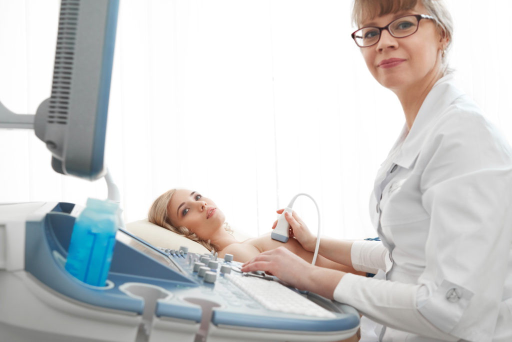
Ultrasounds are performed either by a skilled technician using a hand-held wand or automatically by a machine, in a technique called automated whole-breast ultrasound (AWBU). Radiologists, physicians who specialize in radiology, review the results. Breast ultrasounds are most commonly performed at general radiology (imaging) centers, specialized women’s radiology centers, and large hospitals.
What Are The Different Types of Ultrasounds?
Breast ultrasounds can be 2D or 3D. Ultrasounds are ordered by doctors as “limited” or “complete.” Complete images are images of the entire breast, areola, and all four quadrants. Limited is a specific area of the breast. “Unilateral” ultrasounds are ultrasounds done to one breast only, and “bilateral” ultrasounds are done to both. Like mammograms, breast ultrasounds can be either screening or diagnostic, the first to look for general issues for asymptomatic patients, and the second to investigate specific symptoms or abnormalities from other tests.
Why Do A Breast Ultrasound?
It’s important to note that that breast ultrasounds are not a replacement for regular mammogram screenings. For most women, mammograms remain the best tool for detecting breast cancer early and breast ultrasounds are most often used as a secondary follow-up imaging technique only after a mammogram has shown abnormalities or inconclusive results.
Breast ultrasounds are sometimes ordered as an alternative to mammogram imaging for pregnant women since ultrasounds do not use radiation. And for women with dense breasts, where mammograms are harder to read, your doctor may order both a mammogram and breast ultrasound at the same time. Breast ultrasounds can also be used during guided needle biopsies to help see the area better, and also to drain fluid from cysts.
New York State Breast Density Law Creates Cancer Screening Awareness
What Can Breast Ultrasounds Detect?
Ultrasounds detect breast masses or lumps, cysts, and calcifications. The details of the imagery will show the shape, size, margin (or, shape of its edges) and location of all of these, which will help your doctor determine whether it’s benign (harmless) or malignant (dangerous, possibly a breast cancer tumor). Breast ultrasounds can sometimes miss microcalcifications, though, which is an early sign of breast cancer and is better seen on mammograms (1). Along with other imagery and exam techniques, breast ultrasounds can help diagnose a variety of diseases and conditions including: breast cancer, mastitis, breast abscesses, infections, lesions, mastopathy, fibroadenoma, breast implant ruptures, fibrocystic changes, and gynecomastia, among others.
Breast Ultrasound Insurance, Referrals & Cost
While mammograms are nearly always covered by insurance these days, it is common for many private medical insurers to not cover breast ultrasounds, despite the fact that they are the most common follow-up test to a mammogram. Notably, the Affordable Care Act (ACA) does not pay for diagnostic breast ultrasounds nor does it include screening ultrasounds under their Women’s Preventative Services Guidelines. Medicare will pay, depending on your risk level, but even so, you will need Medicare Part B as well as pay a 20% copay. Paying the full out-of-pocket cost (also called “cash price”), breast ultrasounds can cost $250-$400 not counting follow-up visits and additional fees. Some clinics offer discounts for paying up front or offer a sliding-scale price based on income. Please reach out to us if you need these resources. If you’re lucky enough to have your breast ultrasound covered by your insurer, a referral from your doctor will be required.
Preparation for A Breast Ultrasound
Good news is that there’s not too much to do before a breast ultrasound. You don’t have to eat or drink differently. You may want to dress in something easily removable since you’ll likely be asked to wear a gown above the waist. Keep in mind, too, that the gel used in the test could possibly get on your clothes. Avoid putting any lotions, powders, or deodorants, on your breasts before the test. And if your breasts are tender from breastfeeding or your menstrual cycle, if possible, consider rescheduling during a less tender time.
What To Expect in a Breast Ultrasound
Once you have your gown on, you will be positioned on your back on an exam table, usually with one of your arms over your head. Those that have already had a mammogram will be pleasantly surprised by how comfortable and not painful the procedure is. The technician will put cool gel on the end of a wand and run it along the breast skin in a pattern, watching the images on the monitor. If it is a Doppler ultrasound, you might hear sounds. You could be asked to shift positions at some point. If your doctor or the technician wants more detail on a specific area, the wand will be applied more firmly in that area so the images are clearer, a technique called “spot compression.” A breast ultrasound takes about 30 minutes. Afterwards the tech will clean the gel off or allow you to clean it off. You can dress immediately afterwards and resume everyday activities.
Get our “Thriver Thursdays” Email
Get all the latest cancer prevention and treatment news plus upcoming survivor programs, straight to your inbox every Thursday. Your privacy is important to us.
Breast Ultrasound Results
Some radiology clinics may be able to give you results after your visit. Almost all will be able to give you a CD of your images and a report which you can keep for your records and share with your doctor. They should also send the results to the doctor that ordered the test.
Remember that just because a breast ultrasound finds an abnormality doesn’t mean it’s cancerous or even malignant. Several harmless conditions show up as abnormalities on breast ultrasounds. If there is an abnormality, you will have the option to do further images, do a biopsy, or do a “wait-and-see” approach, where you follow-up with another breast ultrasound in a specific time frame, such as 6 months, to see if the abnormality has changed.
While breast ultrasounds are accurate, and especially useful for young symptomatic women, they are not the primary way to detect breast cancer and work best in tandem with other screening tools like mammograms, breast MRIs, and clinical breast exams. When coupled with mammograms in one study, breast ultrasounds help detect more breast cancer, but also resulted in substantially more false-positives, falsely diagnosing patients with cancer, which resulted in unnecessary biopsies (2). As with most medical procedures, make sure to talk to your doctor about your breast cancer risk levels and which screening methods are a good fit for you.
Many people do not have medical insurance, are under-insured, or are paying for medical treatments out of pocket. Even those that do have insurance coverage can face obstacles to access, something particularly prevalent in communities of color, where Black women are diagnosed at later, less-treatable stages than any other race. Helping everyone in our community get access to vital cancer screenings is the core of our mission at SHAREing & CAREing. If you need help paying for cancer services such as breast ultrasounds, finding providers, communicating in your language, dealing with insurance, or transporting yourself to your appointments, please contact us. We can provide a volunteer patient navigator to help you with your situation and our services are free of charge to those in need.
Sources
- “Ultrasound examination of breast microcalcifications: luxury or necessity?” European Journal of Radiology, 2006
- “Combined Screening With Ultrasound and Mammography vs Mammography Alone in Women at Elevated Risk of Breast Cancer,” The Journal of the American Medical Association, 2008

